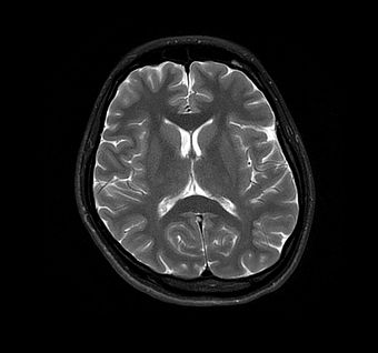
39 brain mri with labels
Brain MRI Atlas on the App Store Brain MRI Atlas is a FREE app that allows you to navigate through hundreds of of labeled brain structures. It is designed for all healthcare professionals as an interactive study and reference tool. Program Features: - Serial sequential axial T2 FLAIR images of the brain. - Structure labels organized by category. Brain lobes - annotated MRI | Radiology Case | Radiopaedia.org 100 public playlists include this case. CT KUB by kazi moin uddin. Learning-SectionalAnatomy by Dr Payam Riahi. Anatomy - Neuro, Head & Neck by Dr Yair Glick . Barin by Dr nermin Nermin Hassan Aboyoussef. 2022 4 by Richard Hodgson. Brain - Anatomy by Dwayne Ian Reading. Anatomy by Mariangela Alvarado Molinaro. Neuro Essentials by Duaa Kanan.
Brain: Atlas of human anatomy with MRI - e-Anatomy - IMAIOS Anatomy of the brain (MRI) - cross-sectional atlas of human anatomy. The module on the anatomy of the brain based on MRI with axial slices was redesigned, having received multiple requests from users for coronal and sagittal slices. The elaboration of this new module, its labeling of more than 524 structures on 379 MRI images in three different views and on 26 anatomical diagrams, took more than 6 months.

Brain mri with labels
Brain MRI: How to read MRI brain scan | Kenhub Reading time: 20 minutes. Normal brain MRI. A brain MRI is one of the most commonly performed techniques of medical imaging. It enables clinicians to focus on various parts of the brain and examine their anatomy and pathology, using different MRI sequences, such as T1w, T2w, or FLAIR. MRI is used to analyze the anatomy of the brain and to ... brain anatomy | MRI coronal brain anatomy | free MRI cross sectional ... This MRI brain coronal cross sectional anatomy tool is absolutely free to use. Use the mouse scroll wheel to move the images up and down alternatively use the tiny arrows (>>) on both side of the image to move the images. Previous. Atlas of BRAIN MRI - W-Radiology The most common MRI sequences used include T1-weighted (T1w) and T2-weighted (T2w) scans (5). T1w sequences display those structures mainly made with fat. Thus, they reveal gray matter as gray, white matter as white, bones as black, and cerebrospinal fluid as black. Meanwhile, T2w sequences highlight structures containing more water.
Brain mri with labels. Subscriptions : NeuromorphoShop, Modeling the Living Human Brain A set of 63 neuroanatomically labeled MRI brain scans Subjects' ages range from 5 to 96 years old. Labels cover the entire brain, are..... more info Max: 1: 2013 Academic Subscription. 30 neuroanatomically labeled MRI brain scans from the ADNI project (part 1) 40 labeled scans from OASIS, 20 subjects scanned twice and labeled twice... Automated MRI image labelling processes 100,000 brain exams in under 30 ... Researchers from the School of Biomedical Engineering & Imaging Sciences at King's College London have automated brain MRI image labeling, needed to teach machine learning image recognition models,... Brain MRI Dataset | Kaggle Brain MRI Dataset | Kaggle. View Active Events. Haşim Mumcu · Updated 3 years ago. arrow_drop_up. 5. New Notebook. file_download Download (8 MiB) more_vert. Functional MRI of the Brain > Fact Sheets > Yale Medicine The exercises increase activity in specific parts of the brain, increasing blood flow and oxygen to them. This activity lights up on the images created by the scanner, giving doctors a visible record of an exact map of the patient's brain. A normal MRI of the brain can last between 20 to 30 minutes, while the fMRI lasts between 40 to 55 minutes.
101 labeled brain images and a consistent human cortical labeling ... To manually label the macroscopic anatomy in magnetic resonance images of 101 healthy participants, we created a new cortical labeling protocol that relies on robust anatomical landmarks and minimal manual edits after initialization with automated labels. MRI Brain Animated Quiz - University of Minnesota Note: spacebar toggles labels; also arrow keys do Previous/Next Sequentially click/tap: first the dot associated with a term; then, its corresponding target dot on the MRI image. If a line connection appears, your choice was correct! Labels · licksylick/Brain-MRI-segmentation · GitHub Brain MRI segmentation using segmentation models. Contribute to licksylick/Brain-MRI-segmentation development by creating an account on GitHub. Labeled imaging anatomy cases | Radiology Reference Article ... This article lists a series of labeled imaging anatomy cases by body region and modality. Brain CT head: non-contrast axial CT head: non-contrast coronal CT head: non-contrast sagittal CT head: angiogram axial CT head: angiogram coronal CT...
What Does a Brain MRI Show? - San Diego Health What does a brain MRI show? The answer is, unfortunately, not very. MRI scans (magnetic resonance imaging) have been around for decades, and the technology has been steadily improving. Today, a brain MRI test can identify whether or not a person has a stroke, or if the person has suffered a traumatic brain injury, or if the person is suffering ... Brain MRI segmentation | Kaggle Journal of Neuro-Oncology, 2017. This dataset contains brain MR images together with manual FLAIR abnormality segmentation masks. The images were obtained from The Cancer Imaging Archive (TCIA). They correspond to 110 patients included in The Cancer Genome Atlas (TCGA) lower-grade glioma collection with at least fluid-attenuated inversion ... CaseStacks.com - MRI Brain Anatomy Labeled scrollable brain MRI covering anatomy with a level of detail appropriate for medical students. Show/Hide Labels. MRI Brain Anatomy. Back to Anatomy overview. Facebook; Twitter; ... Labelled radiographs and CT/MRI series teaching anatomy with a level of detail appropriate for medical students and junior residents. Pelvis. Pelvic MRI anatomy A normative spatiotemporal MRI atlas of the fetal brain for automatic ... Step 3: Fetal brain MRI labels were manually defined and propagated in iterations from the higher GAs to the lower GAs. Step 4: Atlases within one week of any query subject were used to generate an initial segmentation of that subject. The initial segmentations of all subjects within one week of the query subject were used to segment that subject.
Cross-sectional anatomy of the brain - e-Anatomy - IMAIOS Axial MRI Atlas of the Brain. Free online atlas with a comprehensive series of T1, contrast-enhanced T1, T2, T2*, FLAIR, Diffusion -weighted axial images from a normal humain brain. Scroll through the images with detailed labeling using our interactive interface. Perfect for clinicians, radiologists and residents reading brain MRI studies.
101 Labeled Brain Images and a Consistent Human Cortical Labeling ... Labeling the macroscopic anatomy of the human brain is instrumental in educating biologists and clinicians, visualizing biomedical data, localizing brain data for identification and comparison, and perhaps most importantly, subdividing brain data for analysis. Labeled anatomical subdivisions of the brain enable one to quantify and report brain imaging data within brain regions, which is routinely done for functional, diffusion, and structural magnetic resonance images (f/d/MRI) and positron ...
Labeled MRI Brain Scans - Neuromorphometrics Labeled MRI Brain Scans. We have the most comprehensively labeled scans and the largest number of scans available anywhere. We label the entire brain and divide the cortex into regions based on gyral and sulcal landmarks using 1) the "General Segmentation" Protocol defined by the MGH Center for Morphometric Analysis (see here ), and 2) the brainCOLOR Cortical Parcellation Protocol (from here ).
MRI Brain Atlas Via a toggle button, either MRI images or approximately comparable Brain Transection images may be viewed with or without labels. Navigation & Labels. The home page presents two menus for locating MRI images per transection level. In the MRI Gallery Menu, tap a reduced image to view a particular transection level.
MR Image Classification for Brain Tumor Texture Based on Pseudo-Label ... MR Image Classification for Brain Tumor Texture Based on Pseudo-Label Learning and Optimized Feature Extraction Comput Math Methods Med. 2022 Apr 4;2022:7746991. doi: 10.1155/2022/7746991. ... First, for the small sample of pituitary tumor MRI image data, the T1 and T2 sequence data are uneven or missing; we used the CycleGAN model to perform ...
Clinical MRI Brain Scans Will Enrich the Adult Changes in Thought Study Data Resource - Memory ...
UCLA Brain Mapping Center - ICBM Template Cortical gyri, subcortical structures and the cerebellum have been delineated from the structural brain template and assigned a unique label. The 3-D set of labels can be imported and registered onto the structural MRI of any individual subject through software like BrainSuite.
MRI anatomy | free MRI axial brain anatomy This MRI brain cross sectional anatomy tool is absolutely free to use. Use the mouse scroll wheel to move the images up and down alternatively use the tiny arrows (>>) on both side of the image to move the images.>>) on both side of the image to move the images.
matlab - Brain MRI Segmentation Using FCM (Labeling) - Stack Overflow Brain MRI Segmentation Using FCM (Labeling) I am doing Brain MRI segmentation using Fuzzy C-Means, The volume image is n slices, and I apply the FCM for each slice, the output is 4 labels per image (Gray Matter, White Matter, CSF and the background), how I can give the same label (Color) for each material for all the slices) I am using matlab.
Researchers automate brain MRI image labellin | EurekAlert! Published in European Radiology, this is the first study allowing researchers to label complex MRI image datasets at scale. The researchers say it would take years to manually perform labelling of ...
NITRC: Manually Labeled MRI Brain Scan Database: Tool/Resource Info This is a continuously growing and improving database of high-quality neuroanatomically labeled MRI brain scans, created not by an algorithm, but by neuroanatomical experts. All results are checked and corrected. Regions of interest include the usual sub-cortical structures (thalamus, caudate, putamen, hippocampus, etc), along with ventricles ...
Atlas of BRAIN MRI - W-Radiology The most common MRI sequences used include T1-weighted (T1w) and T2-weighted (T2w) scans (5). T1w sequences display those structures mainly made with fat. Thus, they reveal gray matter as gray, white matter as white, bones as black, and cerebrospinal fluid as black. Meanwhile, T2w sequences highlight structures containing more water.

MRI Brain Activity. Ever hear we use only 10% of brain? Common MYTH used by writers, psychics to ...
brain anatomy | MRI coronal brain anatomy | free MRI cross sectional ... This MRI brain coronal cross sectional anatomy tool is absolutely free to use. Use the mouse scroll wheel to move the images up and down alternatively use the tiny arrows (>>) on both side of the image to move the images. Previous.
Brain MRI: How to read MRI brain scan | Kenhub Reading time: 20 minutes. Normal brain MRI. A brain MRI is one of the most commonly performed techniques of medical imaging. It enables clinicians to focus on various parts of the brain and examine their anatomy and pathology, using different MRI sequences, such as T1w, T2w, or FLAIR. MRI is used to analyze the anatomy of the brain and to ...


Post a Comment for "39 brain mri with labels"